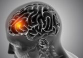
MRI brain scan of children with Tourette’s syndrome may provide clues to disorder pathology
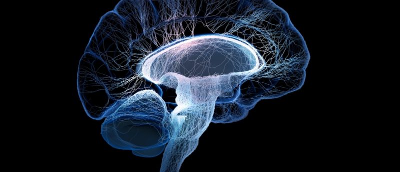
Researchers at Washington University School of Medicine (St. Louis, MO, USA) have utilized structural MRI to identify areas in the brains of children with Tourette’s syndrome that appear markedly different when compared with the same areas in children without the neuropsychiatric disorder, potentially revealing new target regions and avenues of study in Tourette’s syndrome. The results of the study, recently published in Molecular Psychiatry, demonstrates evidence for abnormal brain structure in children and youth with Tourette’s syndrome, consistent with previous findings. “In this study, we found changes primarily in brain regions connected to sensation and sensory processing,” commented co-principal investigator...